UC Merced’s compact campus includes four shared user facilities: the Imaging Microscopy Facility, the Environmental Analytical Lab, the Molecular Characterization Facility, and the Stem Cell Instrument Foundry.
This facility provides the tools for device fabrication such as a contact aligner, atomic layer deposition and profilometer.
The Stem Cell Instrumentation Foundry (SCIF) will provide advanced instruments, student training in cutting-edge techniques, and access to microfabrication collaborators. Selected six graduate scholars will be working in the Stem Cell Instrumentation Foundry’s (SCIF) cleanroom and cell culture labs for up to two weeks period and will primarily get trained on the photolithography and cell culture equipment. SCIF features are as follows:
The newly opened SCIF at UC Merced provides stem cell researchers at UC Merced and throughout California access to advanced instruments, techniques and collaborators for single cell analysis. The SCIF is housed on the main UC Merced campus in a 5420 square-foot facility which includes Class 1000 and 100 clean rooms for micro/nano fabrication, facilities for human and mouse stem cell culture, quantitative cell imagining, and workstations. The SCIF features clean-rooms for micro/nano fabrication and has essential fabrication equipment, providing a broad range of processing capabilities, including facilities for human and mouse stem cell culture and quantitative cell imaging. The facility is equipped with facilities for human and mouse stem cell culture, quantitative cell imaging, and workstations, Nikon TE-2000U inverted epi-fluorescence microscope with CoolSnap HQ2 camera, fluorescent microscopy and fluorescence cell scanning and sorting (FACS), a Laser Scanning Confocal Microscope, FACS cell sorters and analyzers (Aria and Guava) and an 11-color FACS scanner (BD LSR II), Focused Ion Beam, photolithography, acid sink, atomic layer deposition, JEOL Environmental SEM (ESEM), 1 standard X- ray generators, small angle unit, an EXAFS unit and equipment for single crystals or powders (from 2k to 2,500 K), lithography (down to 1.2 μm), vapor deposition systems, reactive ion etching and UV- ozonation tool, and wet chemical processing. Researchers also have access to a wide range of offerings enabling them to rapidly adopt groundbreaking research technologies, connect to online support, workshops and collaborators, conduct computational biology and make new discoveries and develop molecular tools and approaches that will be broadly applicable for basic and applied science and regenerative medicine.
The SCIF is particularly unique because it provides a range of microfluidic-based systems enabling researchers, with no prior knowledge of micro/nano techniques, to custom design devices online to their specific needs, rapidly adopting cutting edge research technologies. Ancillary core services include materials characterization, a vivarium, and computational biology.
SCIF equipment, instruments and services include Cell Culture, Flow Cytometry and Cell Sorting, Microscopy, and Clean Room and Microfabrication. The SCIF is equipped with a variety of resources to provide these services, including: 4 tissue culture hoods, 4 incubators, Flow cytometers, a Nikon Eclipse Ti C1 Confocal System, an NT-MDT NEXT Atomic Force Microscope, a Nikon Eclipse TS-100 Inverted Fluorescent Microscope, Pathology Devices, a Live Cell Stage Top Incubation System, a Bruker Dektak XT Profilometer, a FIB 800x Ion Beam Microscope, a Mask Aligner, a Custom Built Solvent Rinse Station, a Wire Bonder, Drying Oven, UV Developer, Electrospinning Station, Atomic Layer Deposition (ALD), Particle Counter, and a DI Water System.
Nanobiofabrication Summer Training Module participants will work in the Stem Cell Instrumentation Foundry’s (SCIF) cleanroom and cell culture labs for up to two weeks period and will become trained on photolithography and cell culture equipment.
The Imaging Microscopy Facility houses advanced imaging instruments such as Zeiss Gemini SEM 500 scanning electron microscope with sub nanometer resolution, JEOL JR-2010 high-resolution transmission electron microscope and X-ray diffractometry (PANalytical X’Pert PRO Theta/Theta X-ray Diffraction) and the Zeiss LSM 880 laser scanning confocal microscope, which is currently the most advanced, state-of-the-art light microscope at UC Merced.
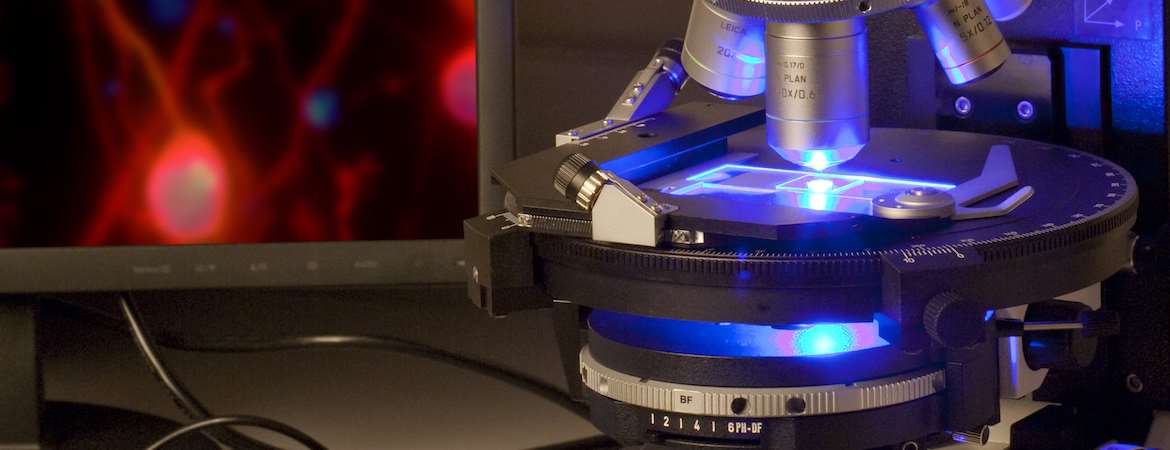
Researchers in physical and biological sciences, as well as engineers, use the lab, which has a particular focus on nanotechnology. Student and faculty researchers find a range of materials characterization techniques, including advanced imaging, elemental analysis and structure determination and can use or be trained in the use of electron microscopes, scanning electron microscopes and transmission electron microscopes (SEM and TEM), atomic force microscope (AFM), small angle X -ray diffraction spectroscopy, raman spectroscopy, atomic force microscopy, dynamic light scattering system.
Molecular Characterization Facility
The Molecular Characterization Facility houses several major instruments, including 400 MHz, 500 MHz and 600 MHz NMR spectrometers, a Horiba Fluorolog 3 fluorimeter, a Bruker Vertex 70 FT-IR spectrometer with a diamond crystal ATR accessory, a high resolution Thermo Electron Exactive Plus LC-MS for detecting small molecules, and a Q Exactive quadrupole Orbitrap LC-MS for high resolution detection of macromolecules.
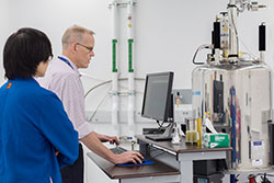
Researchers can search for major and trace elements, selected chemical species and nutrients and organic compounds.
University of California, Davis
UC Merced faculty have access to a 800 MHz Avance III Spectrometer located on the campus of the University of California, Davis, approximately a 2-hour drive north of Merced, CA. The facility is equipped with with a Bruker TCI cryoprobe that provides about a 4:1 Signal-to-Noise improvement over conventional probes. It is highly suited for metabolomic studies, the study of complex biomolecules like proteins and nucleic acids, and routine 1H and 13C NMR experiments on small molecules.
SLAC National Accelerator Laboratory
The Stanford Synchrotron Radiation Lightsource (SSRL) is a multi-program national laboratory exploring frontier questions in photon science, astrophysics, biochemistry, material science, particle physics and accelerator research, and provides synchrotron radiation, a name given to X-rays or light produced by electrons circulating in a storage. These extremely bright x-rays serve as a resource for researchers to perform studies at the atomic and molecular level. Synchrotron radiation laboratories are also used for x- ray scattering and diffraction experiments. SSRL also provides unique educational experiences and serves as a vital training ground for future generations of scientists and engineers.

Lawrence Berkeley National Laboratory
The Molecular Foundry, which houses the state-of-art imaging, synthetic, characterization and modeling capability supports major instrumentation such as, AB SCIEX TF4800 MALDI TOF-TOF Mass Spectrometer, Bruker Avance II 500 MHZ NMR Spectrometer, Lakeshore CPX-HF Superconducting Magnet-Based Horizontal Field Cryogenic Probe Station, Photovoltaic Test Station, Bruker MICROTOF- Q Mass Spectrometer, NanoLog Spectrofluorometer, MBRAUN Thermal Evaporator and VEECO DEKTAK 150 Profilometer and JEOL 2100-F 200 kV Field-Emission Analytical Transmission Electron Microscope and Zeiss Libra 120 Cryo-TEM and Zeiss Gemini Ultra-55 Analytical Field Emission Scanning Electron Microscope, PHI 5400 X-ray Photoelectron Spectroscopy (XPS) System, and Lorentz TEM for high-resolution electron microscopy with in situ XRD capabilities and magnetic field for structural and functional characterizations.


